CT Scan vs MRI: Differences, Uses & Which is Better
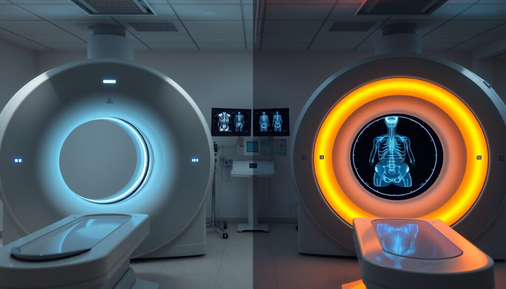
CT scans and MRIs both generate detailed cross-sectional images, but their underlying technologies, clinical strengths, and safety profiles diverge sharply. A recent study at a major cancer center found MRI detected brain abnormalities in 12.6% of cancer patients with neurological symptoms, compared with just 5.2% for CT—a gap that matters when the stakes are high.
Radiation Exposure: CT uses ionizing radiation; MRI uses magnetic fields, none · Speed: CT takes about 10 minutes; MRI takes 45–60 minutes · Soft Tissue Detail: MRI delivers superior contrast resolution; CT excels at bone and spatial resolution · Safety: Both are safe and noninvasive
Quick snapshot
- Exact radiation dose varies by machine model and protocol
- Cost differences fluctuate by region, facility type, and insurance plan
- MRI adoption likely to grow as cost gaps narrow and reimbursement models shift
- Continued debate around appropriate CT use thresholds given radiation concerns
| Factor | CT Scan | MRI |
|---|---|---|
| Primary Use | Trauma, cancer staging, bone imaging | Neurology, musculoskeletal, soft tissue tumors |
| Patient Experience | Open scanner; no tunnel | Enclosed tunnel; some claustrophobia risk |
| Safety | Uses ionizing radiation | No radiation; magnetic fields only |
| Speed | About 10 minutes | 45–60 minutes |
| Cost ( uninsured) | $500–$3,000 | $1,200–$4,000 |
| Soft Tissue Detail | Moderate contrast resolution | Superior contrast resolution |
Which is better MRI or CT scan?
There is no universal answer to which is “better”—it depends entirely on what your doctor is looking for. Each imaging method has distinct strengths, and the choice comes down to the clinical question at hand. According to experts at Memorial Sloan Kettering Cancer Center, both are safe and noninvasive, but they answer different diagnostic questions.
When CT excels over MRI
CT scans win on speed and accessibility. A CT takes roughly 10 minutes, compared with 45–60 minutes for an MRI, making it the preferred choice in emergency rooms when doctors need answers fast. CT also handles patient movement better, which matters for trauma patients who cannot stay still. For bone injuries and acute conditions, CT provides excellent spatial resolution, giving doctors a clear picture of fractures and structural damage. The American College of Radiology advises that no CT imaging be done unless there is clear medical benefit due to radiation exposure, but when that threshold is met, CT delivers rapid, actionable results.
When MRI outperforms CT
MRI’s advantage lies in soft tissue contrast. The technology excels at differentiating between healthy tissue and tumors in the brain, liver, and reproductive organs—areas where CT often struggles for clarity. A study published in PMC and analyzing 170 patients found MRI achieved 78.82% sensitivity for detecting small hepatocellular carcinoma, compared with just 62.35% for CT. For cancer patients presenting neurological symptoms, MRI detected critical brain abnormalities in 12.6% of cases versus only 5.2% for CT in a study of 1,900 patients. That gap can be the difference between finding a metastasis and missing it.
Factors doctors consider
Radiation exposure sits at the top of the list. CT uses ionizing radiation from X-rays, which can cause cell damage potentially leading to cancer if accumulated over time. MRI relies entirely on magnetic fields and radio waves, making it safer for patients who need repeated imaging—particularly cancer patients undergoing monitoring. Doctors also weigh cost (MRI typically runs $1,200–$4,000 versus $500–$3,000 for CT), patient tolerance (claustrophobia affects MRI candidates), and metal implants (which rule out MRI due to its strong magnets).
What does an MRI scan show that a CT scan does not?
MRI reveals soft tissue details that CT simply cannot capture with the same precision. The difference comes down to how the two technologies build their images—MRI maps water molecules in tissues using magnetic fields, while CT relies on X-ray absorption differences between tissue types.
Soft tissue advantages
MRI produces superior contrast resolution for soft tissues, making it the go-to choice for evaluating tumors in the brain, liver, prostate, uterus, and other organs with high water content. For small hepatocellular carcinoma specifically, MRI reached 78.67% diagnostic accuracy versus 67.33% for CT in a peer-reviewed study of 170 patients. The technology also excels at detecting early-stage cancers where treatment is most effective, and at distinguishing between benign and malignant masses based on how tissues appear under different imaging protocols.
Nerve and brain specifics
Nerve damage and brain abnormalities are MRI territory. The technology can visualize spinal cord tissue, nerve roots, and subtle brain lesions that CT cannot adequately assess. For cancer patients with suspected brain metastases, MRI detected critical abnormalities in 12.6% of cases versus 5.2% for CT—a finding with obvious implications for treatment planning. MRI with Diffusion-Weighted Imaging also offers superior diagnostic accuracy for colorectal liver metastases, outperforming contrast-enhanced CT in head-to-head studies.
What Does a CT Scan Show That an MRI Cannot?
CT holds its own in several important areas where MRI falls short. The technology’s speed and bone visualization make it indispensable for certain clinical scenarios—particularly emergencies and structural assessments.
Bone and acute injury detection
CT provides superior spatial resolution for bones and complex anatomical structures. Emergency physicians rely on CT to quickly rule out life-threatening conditions like brain hemorrhage, aortic dissection, and pulmonary embolism. The technology captures cross-sectional images rapidly, making it ideal when seconds matter. For orthopedic applications—evaluating complex fractures, spinal alignment, or bone tumors—CT often delivers the clarity doctors need without the time investment MRI requires.
Cancer detection examples
CT detects many solid tumors and cancers effectively, particularly those with significant structural mass or calcification. Lung cancers, kidney cancers, and bone metastases often show up clearly on CT. The technology excels at cancer staging by revealing tumor size, lymph node involvement, and distant spread. For patients who cannot undergo MRI due to implants or claustrophobia, CT provides a practical alternative that still delivers actionable diagnostic information.
Why do doctors prefer CT scan over MRI?
Doctors reach for CT when clinical urgency, logistics, or patient factors make it the more practical choice. Speed is the biggest driver—CT delivers results in minutes while MRI requires patient commitment to a lengthy, motionless session inside a tunnel.
Speed and availability
CT scanners are more widely available in hospitals and imaging centers, and they produce results faster. For emergency departments, that speed translates directly to patient outcomes. A trauma patient with internal bleeding cannot wait 45 minutes in an MRI tube. CT also handles patient movement better, which matters when dealing with unconscious or agitated patients. According to Duke Tisch Brain Tumor Center, CT’s speed advantage makes it better for emergencies where quick decisions are essential.
Cost and safety factors
MRI typically costs roughly twice as much as CT—$1,200–$4,000 versus $500–$3,000 without insurance. For patients with metal implants, pacemakers, or other ferromagnetic devices, CT is the only safe option since MRI’s strong magnets can cause these devices to malfunction or heat. Claustrophobia affects a significant portion of the population, making open CT scanners more tolerable than MRI’s enclosed tunnel for anxious patients. The American College of Radiology advises minimizing unnecessary CT imaging due to radiation concerns, but when medical benefit clearly outweighs that small risk, CT remains a valuable tool.
Can you see a tumor on a CT scan?
Yes, CT detects many tumors and cancers effectively—but with important caveats. The technology is a cornerstone of cancer staging, helping doctors determine tumor size, lymph node involvement, and whether cancer has spread to distant sites.
Cancer types detected
CT reliably shows lung cancers, kidney cancers, bladder cancers, and bone metastases. The cross-sectional images reveal mass size and location, enabling treatment planning and monitoring. For abdominal organs, CT with contrast can differentiate many tumors from surrounding tissue. For colorectal liver metastases, a PMC cost-effectiveness analysis found contrast-enhanced CT costs $42,874.02 with 8.47 quality-adjusted life years, but MRI with Diffusion-Weighted Imaging still dominated on diagnostic accuracy and long-term value.
Limitations for some cancers
CT struggles with small soft tissue tumors, particularly in organs like the liver and brain. For small hepatocellular carcinoma, CT achieved only 62.35% sensitivity versus 78.82% for MRI in a study of 170 patients. Early-stage tumors that have not yet accumulated significant mass often escape CT detection. Brain tumors near the skull or in areas with complex anatomy can similarly be missed. For these cases, MRI’s superior contrast resolution makes it the preferred follow-up or primary imaging choice.
CT Scan vs MRI: Head-to-Head Comparison
Three clinical metrics stand out when comparing CT and MRI directly: soft tissue sensitivity, cost-effectiveness, and safety profile. The data favors MRI for cancer detection, but CT retains practical advantages in speed and accessibility.
| Metric | CT Scan | MRI | Source |
|---|---|---|---|
| Small HCC sensitivity | 62.35% | 78.82% | PMC (clinical study) |
| Small HCC accuracy | 67.33% | 78.67% | PMC (clinical study) |
| CRLM cost-effectiveness | $42,874.02 / 8.47 QALYs | $40,863.65 / 8.50 QALYs | PMC (cost analysis) |
| Cost-effective at $100K WTP | 6.16% of simulations | 82.99% of simulations | PMC (50,000 simulations) |
| Brain abnormality detection | 5.2% of cases | 12.6% of cases | Hirschfeld Oncology |
| Radiation exposure | Ionizing radiation | None | MSKCC |
| Scan time | ~10 minutes | 45–60 minutes | Docpanel |
MRI outperformed CT across every major cancer detection metric in peer-reviewed studies—higher sensitivity for small tumors, better accuracy for liver metastases, and superior cost-effectiveness in 82.99% of 50,000 Monte Carlo simulations. The gap is not close.
Detailed Specifications and Safety Considerations
| Specification | CT Scan | MRI |
|---|---|---|
| Technology | X-ray ionization | Magnetic fields + radio waves |
| Radiation | Yes — low dose per scan | No |
| Typical scan duration | 5–15 minutes | 30–60 minutes |
| Contrast agents | Iodine-based (kidney/allergy risk) | Gadolinium-based (NSF risk) |
| Claustrophobia risk | Low (open scanner) | Moderate to high (tunnel) |
| Metal implant compatibility | Safe | Contraindicated for ferromagnetic |
| Pregnancy safety | Avoid in first trimester if possible | Generally safe |
| Best for | Bones, acute trauma, fast staging | Soft tissues, nerves, repeated imaging |
| Typical cost (uninsured) | $500–$3,000 | $1,200–$4,000 |
| Average cost | ~$1,200 | ~$2,000 |
Insurance typically covers most costs for medically necessary scans, leaving only a copay—but uninsured patients face the full range. Wisconsin hospital MRI can reach $6,400 due to monopolies, while independent centers offer $650–$850. Geographic and facility-type variation is substantial.
Upsides
- CT provides rapid results in emergencies
- CT costs less and is more widely available
- CT is safe for patients with metal implants
- MRI delivers superior soft tissue contrast
- MRI involves no ionizing radiation
- MRI detects smaller tumors missed by CT
Downsides
- CT radiation accumulates with repeated scans
- CT struggles with subtle soft tissue details
- MRI costs roughly twice as much as CT
- MRI takes 4–6× longer per session
- MRI causes claustrophobia for many patients
- MRI is unsafe for ferromagnetic implant patients
What Experts Say
“The most significant safety distinction between CT and MRI is that an MRI uses no ionizing radiation.”
— Hirschfeld Oncology
“As a precaution, the American College of Radiology advises that no CT imaging be done unless there is a clear medical benefit.”
— Docpanel radiology experts
“MRI was determined to be the dominant strategy” in cost-effectiveness modeling for colorectal liver metastases.
— PMC Study Authors
CT contrast agents carry risks for patients with kidney disease or iodine allergies. Gadolinium used in MRI, while generally safer, has been associated with nephrogenic systemic fibrosis in rare cases. Always disclose your full medical history before any contrast-enhanced scan.
Key Takeaways
CT and MRI answer different diagnostic questions, and the choice is not about which technology is “better” overall—it is about which fits your specific clinical situation. For emergencies and bone imaging, CT’s speed and spatial resolution make it the practical choice. For soft tissue evaluation, tumor detection, and patients needing repeated imaging, MRI’s superior contrast resolution and radiation-free design give it the edge. The evidence from peer-reviewed studies—including MRI’s 78.82% sensitivity for small liver cancers versus 62.35% for CT, and its dominance in 82.99% of cost-effectiveness simulations—suggests MRI should be the default choice whenever soft tissue detail is the priority and patient factors allow.
The American College of Radiology urges minimizing unnecessary CT imaging due to radiation concerns, but that guidance applies to screening use, not medically indicated diagnostic scans. For cancer patients, the trade-off between CT’s speed and MRI’s accuracy is a clinical decision your care team makes based on tumor type, staging goals, and your individual risk profile. Understanding these trade-offs helps you have a more informed conversation with your doctor about which scan makes sense for your situation.
Related reading: ALT blood test · What is a fever
aflac.com, resources.healthgrades.com, myssmi.com, medicinenet.com, tischbraintumorcenter.duke.edu, milleniummri.com
While CT scans often prove cheaper than MRIs, providers like those in Pacific Radiology price list reveal specific costs varying by location and procedure.
Frequently asked questions
CT scan vs MRI: which is safer?
MRI is safer for frequent imaging since it uses no ionizing radiation. CT involves low-dose radiation that accumulates over time, raising cancer risk with repeated scans. The American College of Radiology advises minimizing unnecessary CT use, but medically indicated CT scans remain appropriate when benefits outweigh risks.
Do you go in a tunnel for a CT scan?
No. CT scanners are open or nearly open, with the patient lying on a table that slides through a wide ring. This makes CT more tolerable for claustrophobic patients. MRI scanners enclose the patient in a tunnel-like tube, which triggers anxiety in many people. Open MRI options exist but may compromise image quality.
What is the most common reason for a CT scan?
Emergency room evaluation is the most common use—CT rapidly rules out strokes, internal bleeding, pulmonary embolism, and major fractures. Cancer staging, trauma assessment, and pre-surgical planning round out the top reasons doctors order CT scans.
MRI or CT scan for nerve damage?
MRI is the clear choice for nerve damage evaluation. The technology visualizes spinal cord tissue and nerve roots with detail CT cannot match. For herniated discs, spinal cord compression, and peripheral nerve tumors, MRI provides the diagnostic clarity doctors need to guide treatment.
What cancers cannot be detected by CT scan?
CT struggles with small soft tissue tumors, particularly small hepatocellular carcinoma, early brain metastases, and some abdominal cancers. MRI’s superior contrast resolution catches tumors CT misses—particularly those involving subtle tissue differences or located in areas with complex surrounding anatomy.
Why did my doctor order an MRI instead of a CT scan?
Your doctor likely needs superior soft tissue detail—perhaps evaluating a tumor’s characteristics, assessing nerve involvement, or monitoring cancer that requires clear visualization of subtle tissue changes. MRI’s lack of radiation also makes it preferable for patients who need repeated imaging over time.
CT scan vs MRI price difference
Without insurance, CT ranges $500–$3,000 while MRI ranges $1,200–$4,000. Average costs sit around $1,200 for CT and $2,000 for MRI. Insurance typically covers most costs for medically necessary scans, leaving only a copay. Regional variation is significant—some Wisconsin hospital MRIs reach $6,400 while independent centers offer $650–$850.
How long does each scan take?
CT takes about 10 minutes total. MRI requires 45–60 minutes, sometimes longer depending on the body area and whether contrast is used. The time difference matters in emergencies and for patients who struggle with lying still.
More related posts
 Maryland Terrapins Men’s Basketball – 2025-26 Roster Preview
 What Is Sugar Alcohol – Benefits, Risks and Uses
 How Long Do Flies Live – Housefly Lifespan Facts (15-30 Days)
 Bad Boys 4 Cast: Full List with Will Smith, Martin Lawrence
 United Airlines Flight Emergency Landing: Recent Incidents
 Lakers vs Orlando Magic Match Player Stats & Box Scores
 Best Feed Store Near Me in Ireland: Top Agri Merchants
 The Count of Monte Cristo – Plot Summary and Character Guide